
Facilities:
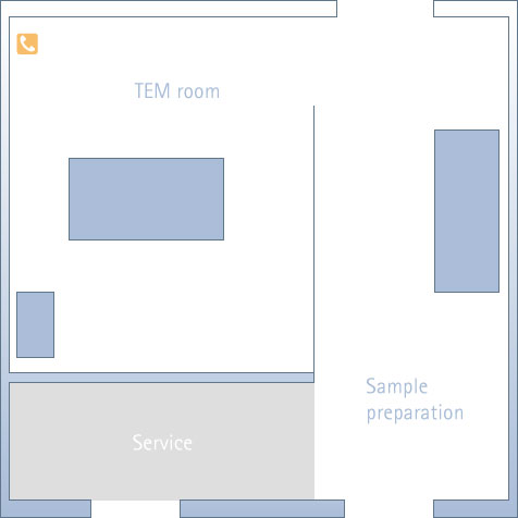
sei in: Map » Ground Floor » Laboratory
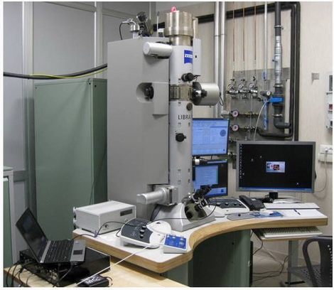
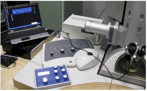
Transmission Electron Microscopy Lab
Equipment
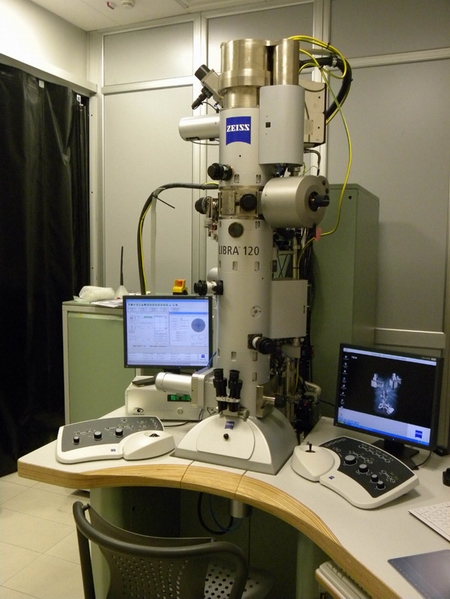
- Zeiss Libra 120 Plus transmission electron microscope: acceleration voltage 60-120 Kv, point-point resolution 0.34 nm in TEM mode and < 1.5nm in STEM mode.
- 16 bit CCD camera 2k x 2k bottom mounted
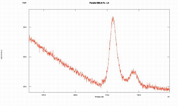
- In column OMEGA spectrometer for EELS and EFTEM imaging
- STEM with High Angle Annular Dark Field (HAADF) detector
- Tomographic single tilt sample holder. This holder can host two grids and allows a maximum tilt of ± 75°
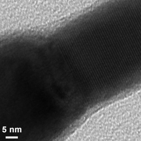
- Conventional and high resolution TEM imaging
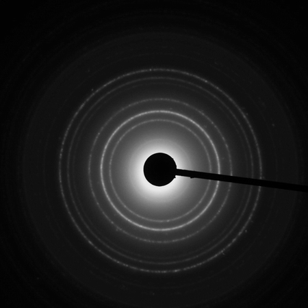
- Electron diffraction with parallel or convergent beam
- Z-contrast STEM imaging with the HAADF detector
- Energy filtered TEM imaging
- Electron Energy Loss Spectrometry EELS
- TEM or STEM electron tomography
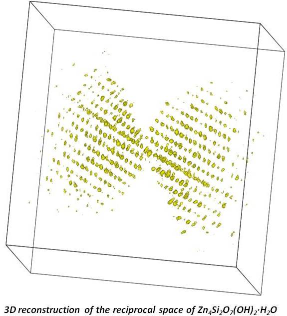
- Nanomegas Digistar P1000 precession unit for collecting precessed electron diffraction patterns, with ADT3D software for three dimensional reconstruction of the reciprocal space and intensity integration.
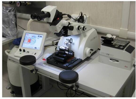
- UC7 Leica Ultramicrotome equipped with the cryo unit FC7 for cutting samples at low temperature down to -185 °C
- Up-right M80 Leica Microscope equipped with CCD color camera for recording images
-
Accessories:
Available techniques:
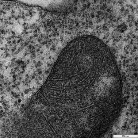
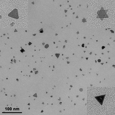
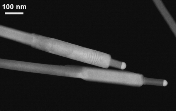
The transmission electron microscope is used to characterize on one side the nanoparticles and the nanostructures (nanowires, eterostructures…) produced in chemistry and CBE-MCVD laboratories and also cell and tissues processed by the biology laboratories at NEST.
© 2009 NEST · Scuola Normale Superiore di Pisa |
W3C quality assurance: xhtml 1.0 strict